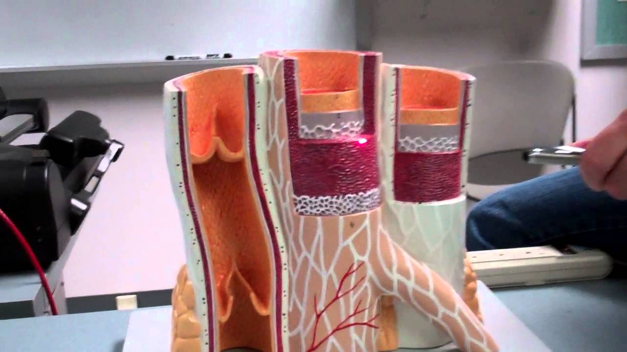
Blood Vessels Labeled : Blood Vessel Histology Model - YouTube

The function and structure of each segment of the peripheral vascular system vary depending on the organ it supplies. Related posts of the human blood vessels labeled digestive system worksheet answers. The thick outermost layer of a vessel (tunica adventitia or tunica externa) is made of connective tissue. When sphincter muscles are relaxed, the capillary bed is open, and blood flows through the capillaries. The major arteries in the body. It extends on each side of the neck and divides at the level of the larynx into two branches: A primary purpose and significant role of the vasculature is its participation in oxygenating the body. Blood vessels consist of arteries, arterioles, capillaries, venules, and veins. Deoxygenated blood from the peripheral veins is transported back to the heart from capillaries, to venules, to veins, to the right side of the heart, and then. Use key choices to identify the blood vessel tunic described. Veins of the head and trunk.
Arteries and veins are composed of three tissue layers. When sphincter muscles are relaxed, the capillary bed is open, and blood flows through the capillaries. Bulky middle tunic contains smooth muscle and elastin 3. Arterioles connect to capillaries, which are the smallest blood vessels and are where the exchange of oxygen, nutrients, and waste occurs between the. Name the blood vessels labeled 'e'. Figure 12.11 anatomy of a capillary bed. The thick outermost layer of a vessel (tunica adventitia or tunica externa) is made of connective tissue.

Deep veins, located in the center of the leg near the leg bones, are enclosed by muscle.
A primary purpose and significant role of the vasculature is its participation in oxygenating the body. Blood is supplied to parts within the neck, head and brain through branches of the subclavian and common carotid arteries. Arteries and veins are composed of three tissue layers. This set is often in folders with. Name the blood vessels labeled 'e'. Deep veins, located in the center of the leg near the leg bones, are enclosed by muscle. Blood vessels are the specially designed tubes that carry blood throughout the body. This video covers one of our blood vessel models The common cartoid artery extends from the brachiocephalic artery. Very small branches that collect the blood from the various organs and parts are called venules, and they unite to form veins, which return the blood to the heart. The upper limb is truly a complex part of the human body.
The smallest arteries are called arterioles. This article lists a series of labeled imaging anatomy cases by system and modality. The major arteries in the body.
A capillary bed forms a maze of capillary vessels that lies between an arteriole and a venule.
Tutorials and quizzes on the circulation of blood and the anatomy, structure, and physiology of blood vessels, using interactive animations and diagrams. Bulky middle tunic contains smooth muscle and elastin 3. Anatomy of a capillary bed. Anatomy of blood vessels review sheet 32 261 microscopic structure of the blood vessels 1. The common cartoid artery extends from the brachiocephalic artery. •formed where capillaries unite • extremely porous 1) venules: You can imagine the aorta and ivc as the two trees, with all. When sphincter muscles are relaxed, the capillary bed is open, and blood flows through the capillaries. Digestive system worksheet answers 12 photos of the digestive system worksheet answers digestive system worksheet answer key, digestive system worksheet pearson education, human digestive system worksheet with answers, the digestive system digestion and absorption worksheet answers, the human digestive. This set is often in folders with. These structures are common sites for conditions that cause narrowing or blockage of the blood vessels.
This set is often in folders with. The vessels that carry blood away from the heart are called arteries, and their very small branches are arterioles. The major veins in the These structures are common sites for conditions that cause narrowing or blockage of the blood vessels. Anatomy of blood vessels review sheet 32 261 microscopic structure of the blood vessels 1. The adventitia or outer layer which provides structural support and shape to the vessel
The major arteries in the body.
Eventually, the smallest arteries, vessels called arterioles, further branch into tiny capillaries, where nutrients and wastes are exchanged, and then combine with other vessels that exit capillaries to form venules, small blood vessels that carry blood to a vein, a larger blood vessel that returns blood to the heart. The thick outermost layer of a vessel (tunica adventitia or tunica externa) is made of connective tissue. Veins of the head and trunk. Veins of the lower body. See more ideas about anatomy, blood vessels anatomy, medical anatomy. A vein is a blood vessel that conducts blood toward the heart. This set is often in folders with. As the abdomen and pelvis contain the majority of internal organs, these regions need to be supplied by an extensive network of arteries and veins. This video covers one of our blood vessel models Anatomy of a capillary bed. The vessels that carry blood away from the heart are called arteries, and their very small branches are arterioles. The common cartoid artery extends from the brachiocephalic artery. Blood vessel labeling online quiz;

Blood vessels consist of arteries, arterioles, capillaries, venules, and veins.

The vessels that carry blood away from the heart are called arteries, and their very small branches are arterioles.

Use key choices to identify the blood vessel tunic described.

See more ideas about anatomy, blood vessels anatomy, medical anatomy.

Vessel networks deliver blood to all tissues in a directed and regulated manner.

Blood is supplied to parts within the neck, head and brain through branches of the subclavian and common carotid arteries.

When sphincter muscles are relaxed, the capillary bed is open, and blood flows through the capillaries.

This video covers one of our blood vessel models
 venules:")
You can imagine the aorta and ivc as the two trees, with all.
 is made of connective tissue.")
When sphincter muscles are relaxed, the capillary bed is open, and blood flows through the capillaries.

Deep veins, located in the center of the leg near the leg bones, are enclosed by muscle.

The function and structure of each segment of the peripheral vascular system vary depending on the organ it supplies.

The function and structure of each segment of the peripheral vascular system vary depending on the organ it supplies.

Microscopic anatomy of blood vessels structure of blood vessels (a) arteries and (b) veins share the same general features, but the walls of arteries are much thicker because of the higher pressure of the blood that flows through them.

Arterioles connect to capillaries, which are the smallest blood vessels and are where the exchange of oxygen, nutrients, and waste occurs between the.

This video covers one of our blood vessel models

The tunic intima of blood vessels, like the endocardium of the heart, is made of simple squamous epithelium.

This video covers one of our blood vessel models

Veins (in blue) are the blood vessels that return blood to the heart.

The tunic intima of blood vessels, like the endocardium of the heart, is made of simple squamous epithelium.

A capillary bed forms a maze of capillary vessels that lies between an arteriole and a venule.

Tutorials and quizzes on the circulation of blood and the anatomy, structure, and physiology of blood vessels, using interactive animations and diagrams.

Anatomy of a capillary bed.

Anatomy of a capillary bed.

Best quiz blood vessel labeling;
 are the blood vessels that return blood to the heart.")
This article lists a series of labeled imaging anatomy cases by system and modality.

The vessels that carry blood away from the heart are called arteries, and their very small branches are arterioles.

The smallest arteries are called arterioles.

The adventitia or outer layer which provides structural support and shape to the vessel

Deoxygenated blood from the peripheral veins is transported back to the heart from capillaries, to venules, to veins, to the right side of the heart, and then.

The major arteries in the body.

Arteries and veins are composed of three tissue layers.

Posting Komentar untuk "Blood Vessels Labeled : Blood Vessel Histology Model - YouTube"